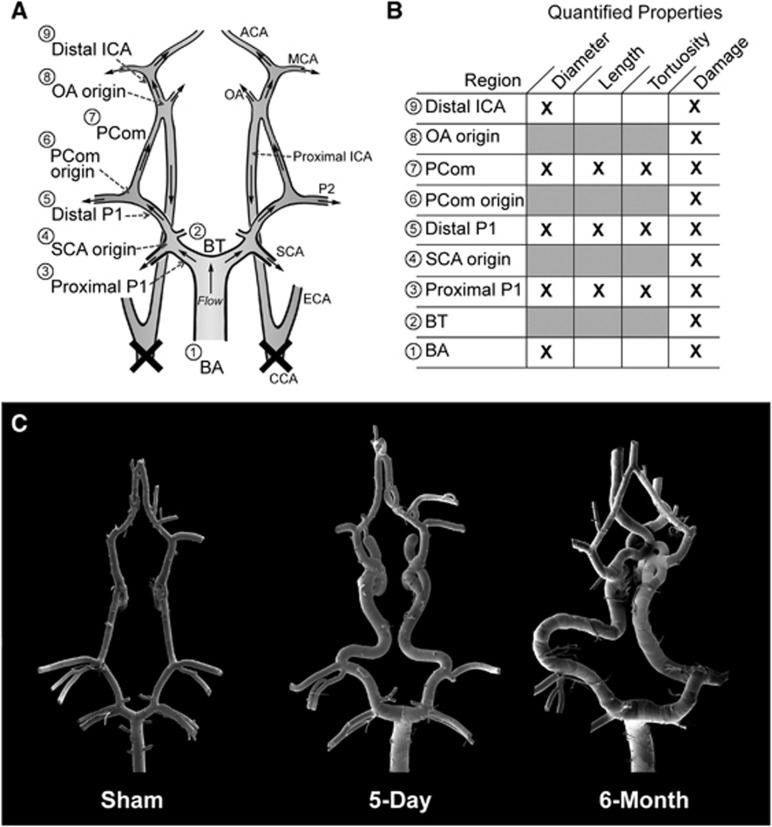
Figure 1.
Overview of morphologic changes that developed in the rabbit circle of Willis (CoW) after bilateral carotid ligation. (A) A schematic showing flow direction after bilateral carotid ligation and nine arterial regions where damage was measured. (B) A chart detailing the quantified properties at each region. (C) Mosaic scanning electron microscopy images of vascular corrosion casts of representative CoW from the sham, 5-day, and 6-month groups. ACA, anterior cerebral artery; BA, basilar artery; BT, basilar terminus; CCA, common carotid arteries; ECA, external carotid artery; ICA, internal carotid artery; MCA, middle cerebral artery; PCom, posterior communicating artery; SCA, superior cerebellar artery.

