Figure 3.
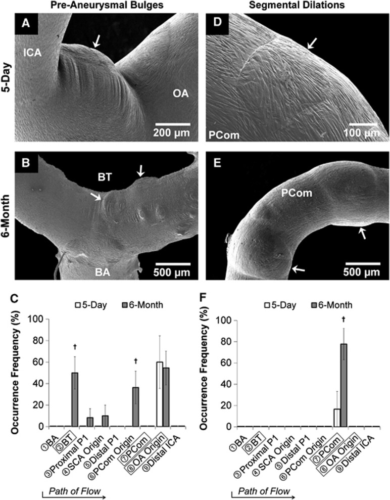
Gross aneurysmal remodeling in the circle of Willis after bilateral carotid ligation evident from scanning electron microscopy images of vascular casts (A, B, D, E) with occurrence frequency at each arterial region being examined (C, F). Left column (A–C): Preaneurysmal bulges. In the 5-day group, preaneurysmal bulges presented only at the ophthalmic artery (OA) origin (A), whereas in the 6-month group notable bulges presented at both the OA origin and the basilar terminus (B), with less frequent bulging at other regions (C). Right column (D–F): Segmental dilations. In the 5-day group, shallow segmental dilations formed on the posterior communicating artery (PCom) (D), whereas multiple large segmental dilations presented on the PCom at 6 months (E, F). (†P<0.05 compared against 5-day). BA, basilar artery; BT, basilar terminus; ICA, internal carotid artery; SCA, superior cerebellar artery.

