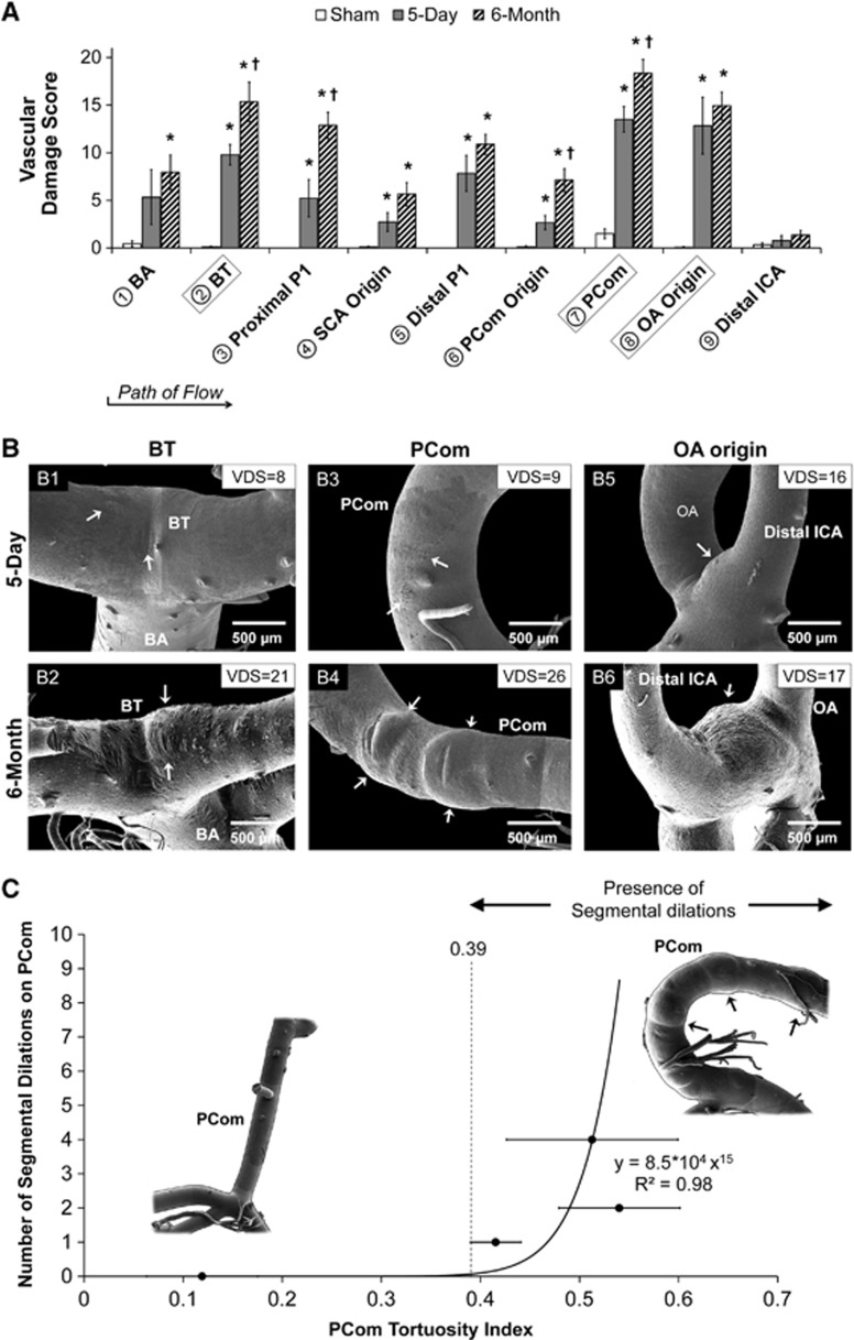
Figure 5.
Quantification of aneurysmal changes in the circle of Willis (CoW) after bilateral carotid ligation. (A) Vascular damage score (VDS), which summates individual values accounting for different types of aneurysmal changes (as defined in Table 1) in each arterial region on the CoW of rabbits from each group, evaluated by 3 masked observers (mean±s.e.). Damage was found throughout the CoW at 5 days and was increased at 6 months in all locations. (*P<0.05 compared against sham. †P<0.05 compared against 5-day). (B–G) Representative scanning electron microscopy images of regions with high scores: (B1) VDS=8 owing to smooth muscle cell imprints (loss of internal elastic lamina, IEL), (B2) VDS=21 owing to preaneurysmal bulging, (B3) VDS=9 because of IEL fenestrations and IEL loss, (B4) VDS=26 owing to segmental dilations, (B5 and 6) VDS are similar (16 and 17, respectively) owing to preaneurysmal bulging. (C) Correlation of the number of segmental dilations and tortuosity index on the posterior communicating artery (PCom). PComs were grouped by number of dilations and the average tortuosity index of each group was calculated (error bars, s.e.). A plot of the number of dilations versus average tortuosity index was fitted to a power function. As the number of dilations is quantized, a tortuosity index threshold of 0.39 is drawn, suggesting that dilations only develop on PComs that are sufficiently tortuous. BA, basilar artery; BT, basilar terminus; ICA, internal carotid artery; OA, ophthalmic artery; SCA, superior cerebellar artery.

