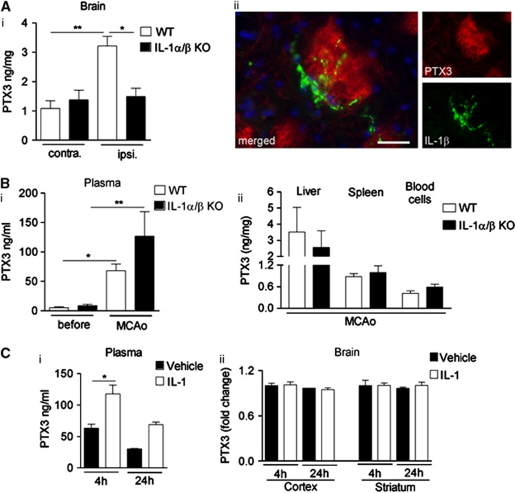
Figure 2.
Central, but not peripheral pentraxin-3 (PTX3) expression is interleukin-1 (IL-1)-dependent after cerebral ischemia. (A) Enzyme-linked immunosorbent assay (ELISA) analyses of brain from wild type (WT) and IL-1α/β knockout (KO) mice 24 hours after middle cerebral artery occlusion (MCAo) indicates that brain PTX3 protein levels are elevated in the ipsilateral hemisphere of WT, but not of IL-1α/β KO mice (i). IL-1β-positive (green) cells are found in close vicinity of PTX3-positive structures (red) within the ipsilateral striatum (ii). (B) Plasma PTX3 increases 24 hours after MCAo in both WT and IL-1α/β KO mice (i), and PTX3 levels in liver, spleen, and blood cells do not vary between genotypes (ii), as measured by ELISA. (C). Peripheral IL-1β injection (i.p.) results in an upregulation of PTX3 in plasma (i), but not in cerebral cortex or striatum (ii). Scale bar, 25 μm. *P<0.05, **P<0.01, one-way analysis of variance followed by Bonferroni's post hoc test (A, B), n=3; (C), i n=3, ii n=2 to 3). Error bars show s.e.m.

