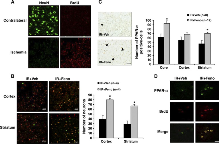
Figure 5.
Evaluation 7 days after the induction of cerebral ischemia of the peroxisome proliferator-activated receptor-α (PPAR-α) agonist fenofibrate effects on neurogenesis in rat. (A) Cell proliferation occurred in the ischemic area as evidenced by a significant increase in the number of cells incorporating bromodeoxyuridine (BrdU) compared with contralateral area of the lesion induced by ischemia. Scale bar, 20 μm. (B) Some of these cells differentiate into neurons, as shown by BrdU and NeuN colabelling in the ischemic area. Fenofibrate administration significantly increased cell proliferation and the number of cells colabelled by BrdU and NeuN both in the striatum and in the cortex; *P<0.05 vs. IR+Veh group. Scale bar, 20 μm. (C) After 7 days of fenofibrate treatment, the number of PPAR-α-expressing cells was also significantly higher in the core of ischemia and in the striatum; *P<0.05 vs. IR+Veh group. (D) Fenofibrate administration significantly increased cell proliferation of cells colabelled by bromodeoxyuridine (BrdU) and PPAR-α. Scale bar, 10 μm.

