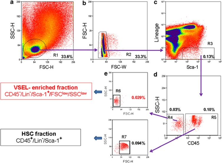
Figure 3.
Classical sorting strategy for murine BM-derived VSEL isolation by fluorescence-activated cell sorting (FACS). Agranular, small events ranging from 2 to 10 μm (as initially set up with Flow Cytometry Size beads, Invitrogen/Molecular Probes) are included in an ‘extended lymphgate' on an FSC vs SSC dot plot (region R1; a). The population of cells from region R1 may be additionally depleted of doublets (gate R2; b) to enhance sorting purity (b). The single-cell fraction from gate R2 is further analyzed for Sca-1 and Lin expression and exclusively Lin−/Sca-1+ cells are gated (R3) to avoid erythroblast contamination (c). The population from region R3 is subsequently separated into CD45− and CD45+ subpopulations visualized in regions R4 and R5, respectively, on a CD45 vs SSC dot plot (d). CD45-dim objects are preferentially included in the CD45+ population (region R5). If BM cells are gated strictly according to these steps (with special caution for R1, R3, R4 and R5 gate set-up), the populations of VSELs and HSCs derived from regions R4 and R5, respectively, separate cleanly when ‘back-gated' on FSC vs SSC dot plots (e). Both stem cell fractions may be additionally purified by final gating, including size-related regions R6 and R7 for VSEL and HSC sorting, respectively. Percentages represent the content of each fraction in the representative murine BM sample.

