
Figure 3. Structure of the active SecY channel.
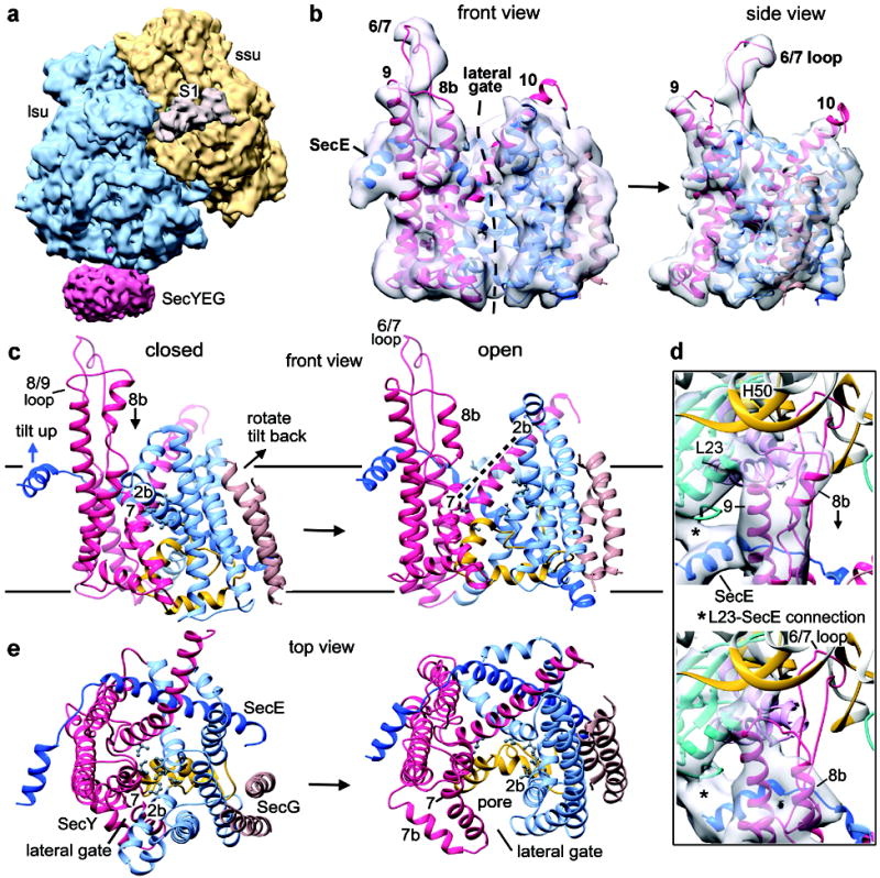
a, Structure of the E. coli RNC–SecY channel complex, with large and small ribosomal subunits in blue and gold, respectively, the SecY complex in red, and ribosomal protein S1 in tan. b, Front and side views of the channel fit into the segmented density map (grey). The nascent chain was omitted for clarity. The N-terminal half of SecY is in light blue, the C-terminal half in red, SecE in dark blue, and SecG in brown. c, Comparison of front views of the closed and open E. coli SecY channels with the approximate position of the membrane indicated by solid horizontal lines. The N-terminal half of SecY is in light blue, the C-terminal half in red, SecE in dark blue, SecG in brown, and the plug in yellow. Some movements during channel opening are indicated, such as the rotation and tilting of the N-terminal half of SecY, the tilting of SecE, and the movement of helix 8b. Labels for helices 2b and 7 are placed at the same position in the closed and open channel. Pore residues forming the constriction in the closed channel are indicated with grey balls and sticks. d, Connections of the ribosome with the 8/9 loop of SecY and the cytoplasmic helix of SecE in the closed and open channels (upper and lower panels, respectively). Note the large movement of helix 8b towards the membrane. e, As in c, but viewed from the top.