Fig. 1.

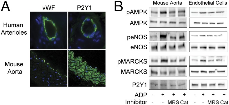
P2Y1 receptor expression and signaling responses in vascular preparations and cultured endothelial cells. A shows representative photomicrographs of human arterioles and murine aortic preparations that were fixed, paraffin-embedded, and stained with antibodies directed against the endothelium-specific marker vWF or the P2Y1 receptor, as indicated. Nuclei were stained with DAPI. Images were obtained by confocal imaging, as discussed in the text. In B, immunoblots were analyzed in murine aortic preparations or cultured endothelial cells that were incubated with ADP (50 μM, 30 min) in the presence or absence of the P2Y1-specific blocker MRS2179 (5 μM) or of the cell permeant H2O2-catabolizing enzyme PEG-catalase (100 U/mL). Membranes were probed with total and phosphospecific antibodies directed against eNOS, AMPK, and MARCKS; P2Y1 served as loading control. Fig. S1 A and B show statistical analyses of pooled data from four identical immunoblot experiments that yielded similar results.
