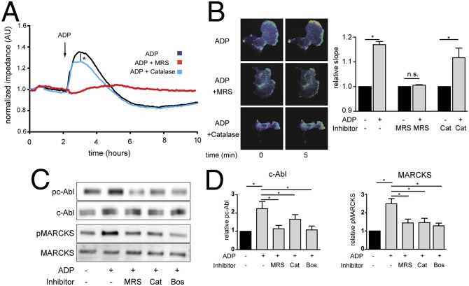
Fig. 2.
ADP- and H2O2-mediated changes in endothelial cell impedance, Rac1 activation, and phosphorylation responses. A shows representative tracings of endothelial cells analyzed in impedance measurements in the presence or absence of ADP (50 μM), the P2Y1 receptor blocker MRS2179 (5 μM), or the H2O2 scavenger PEG-catalase (100 U/mL). The findings shown are representative of three identical experiments that yielded similar results. B shows representative photomicrographs of endothelial cells transfected with a plasmid encoding a Rac1 FRET biosensor and then analyzed by quantitative time-lapse microscopy before and 5 min after the addition of ADP in the presence or absence of MRS2179 (MRS) or PEG-catalase (Cat); pooled data are shown from four identical experiments, presenting the slope of the fluorescence increase following the addition of ADP, measured 5 min after adding ADP in the presence or absence of MRS2179 or PEG-catalase. C shows representative immunoblots of cultured endothelial cells incubated with ADP in the presence or absence of MRS2179, the c-Abl inhibitor bosutinib, or PEG-catalase as indicated, probed with antibodies as shown. D shows statistical analyses of pooled data from three identical experiments that yielded similar results; *P < 0.05 (ANOVA).

