Fig. 3.
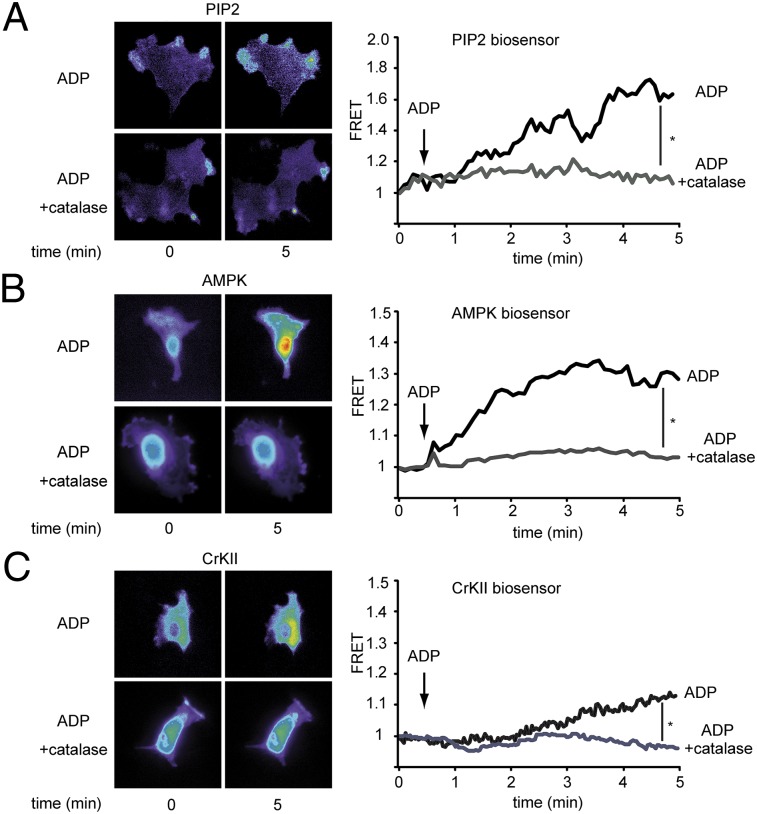
Effects of PEG-catalase on ADP-dependent modulation of PIP2, AMPK, and c-Abl. Endothelial cells were transfected with plasmids encoding FRET biosensors specific for PIP2 (A), AMPK (B), and CrKII (C) and then analyzed by quantitative FRET microscopy before and after the addition of ADP (50 μM, 5 min) in the presence and absence of PEG-catalase. A–C Left show representative photomicrographs; A–C Right show representative tracings of FRET ratios as well as statistical analysis of the ADP-promoted FRET slope change, as pooled and plotted from four independent experiments; *P < 0.05 (ANOVA).

