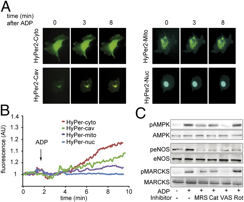
Fig. 5.
Detection of ADP-promoted changes in intracellular H2O2 levels. A shows representative photomicrographs of endothelial cells transfected with differentially targeted variants of the hydrogen peroxide specific biosensor HyPer2 expressed in cytosol (Cyto), caveolae (Cav), nucleus (Nuc), or mitochondria (Mito), as indicated, and then treated with ADP. B shows representative tracings for the individual differentially targeted HyPer2 constructs; ADP addition is indicated by an arrow. C shows immunoblots of cultured endothelial cells incubated with ADP in the presence or absence of MRS2179 (“MRS”; 5 μM), PEG-catalase (“Cat”; 100 U/mL), the NADPH oxidase inhibitor VAS2870 (“VAS”; 10 μM), or the inhibitor of mitochondrial respiration rotenone (“Rot”; 10 μM) as indicated. Membranes were probed with total and phosphospecific antibodies for AMPK, eNOS, and MARCKS. The results shown are representative of three identical experiments that yielded similar results; statistical analyses of pooled data are presented in Fig. S1C.

