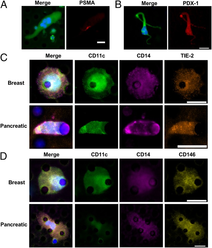
Fig. 3.
CAMLs stain for monocytic, endothelial, and specific tissue markers. (A) CAML from a prostate cancer patient stained with DAPI (blue) and anti–cytokeratins-FITC (green) (Left) and with anti–PSMA-Dylight594 (red) (Right). (B) CAML from a pancreatic cancer patient stained with DAPI (blue) and anti–cytokeratins-FITC (green) (Left) and anti–PDX-1-Dylight594 (red) (Right). (C) CAMLs from samples from patients with breast (Upper Row) or pancreatic (Lower Row) cancer. Images from left to right show a merged image with DAPI, anti-CD11c (monocyte marker), anti-CD14 (monocyte marker), and anti-TIE-2 (angiogenic marker) staining and images of staining for individual markers. (D) CAMLs from samples from patients with breast (Upper Row) or pancreatic (Lower Row) cancer. Images from left to right show a merged image with DAPI, anti-CD11c, anti-CD14, and anti-CD146 (endothelial marker) staining and images of staining for individual markers. (Scale bars, 20 µm.)

