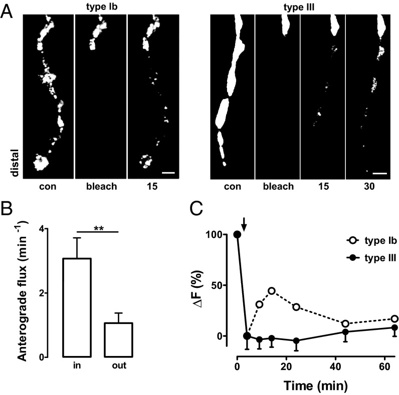
Fig. 3.
DCV capture in type III boutons is efficient. (A) Time-lapse images of type Ib and type III boutons before and after photobleaching boutons. FRAP shows DCV accumulation is marked in the most distal type Ib boutons and the most proximal type III boutons. Contrast was adjusted to visualize puncta. Numbers indicate time in minutes. Proximal unbleached boutons are shown at the top of images. (Scale bar, 2 μm.) (B) DCV anterograde flux into and out of photobleached proximal type III boutons shows high capture efficiency in type III boutons (n = 7). **P < 0.01. (C) Change in neuropeptide content after a 3-min depolarization (indicated by arrow) of type III boutons (black circles) (n = 8). Data are normalized to the initial decrease in fluorescence (ΔF). Dotted curve (open circles) shows the rebound in type Ib boutons (replotted from ref. 17) evoked by the same stimulus that is indicative of activity-dependent DCV capture.

