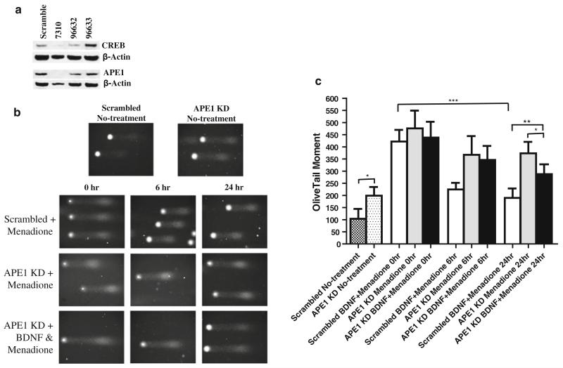
Fig. 4.
Posttranscriptional suppression of APE1 production blocks the ability of BDNF to enhance repair of oxidative DNA damage in cultured cerebral cortical neurons. a Cultured cortical neurons were infected with a lentiviral vector that expresses an shRNA directed against the CREB mRNA, and 3 days later, cells were harvested for immunoblot analysis. The upper band is CREB and that the numbers, 7310, 96632, and 96633, are the three different shRNAs directed against the CREB mRNA. Neurons infected with the CREB shRNA exhibited greatly reduced levels of APE1 compared to parallel cultures infected with a lentivirus to express a shRNA with a scrambled sequence. Similar results were obtained in 3 separate experiments. b Representative images of neuronal nuclear DNA as visualized in comet assay gels. The upper two images show nuclei from cortical neurons in cultures infected with lentiviruses to express either scrambled shRNA or APE1 shRNA. The next three rows of images show nuclear DNA from cortical neurons that had been treated with 20 μM menadione for the indicated time periods. Prior to exposure to menadione, the neurons were infected with lentiviruses to express scrambled shRNA or APE1 shRNA, in the absence or presence of 10 ng/ml BDNF. c Results of quantification of comet assay results. Values are the mean and SD (n = 4–6 separate cultures). *P < 0.05, **P < 0.01, ***P < 0.001

