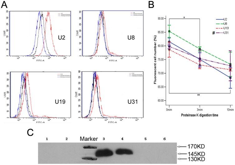
Figure 3. Aptamers U2 and U8 target EGFRvIII specifically.
(A) As protease K digestion time increasing from 3 min (black) to 10 min (blue), transition of the fluorescence intensity shift to the left illustrated a signal intensity decrease. The binding of FITC-labeled aptamers to intact U87-EGFRvIII cells (0 min) was used as positive control (red). (B) Changes in cell number with fluorescence as digestion time increasing. Data represent mean ± SD of three independent experiments. * P<0.05, ** P<0.01. #: There was no statistical difference at different time points in U31 group. (C) The specific interaction of U2 and U8 with EGFRvIII was determined by affinity purification on streptavidin beads of cell lysate treated with biotin-labeled aptamers followed by immunoblotting with anti-EGFR antibody. EGFRvIII has a molecular weight of 145 kDa compared with that of 170 kDa for EGFRwt. Obvious bands at 145 kDa indicated the spicific target of aptamers was EGFRvIII. Lanes (left to right): 1, GN with U87MG cell lysate; 2, GN with U87-EGFRvIII cell lysate; 3, U2 with U87-EGFRvIII cell lysate; 4, U8 with U87-EGFRvIII cell lysate; 5, U2 with U87MG cell lysate; 6, U8 with U87MG cell lysate. Three independent experiments were performed.

