
Figure 1. Hematoxylin and eosin (H&E) staining of wounded WT and FMOD-null adult mice skin.
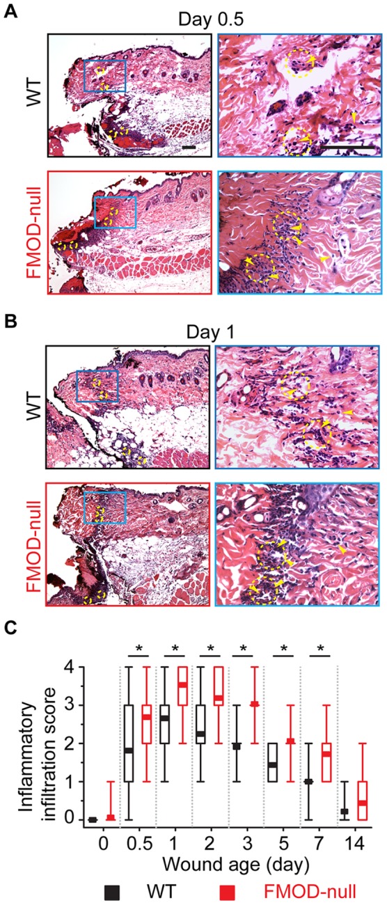
(A) At day 0.5 post-injury, minimal (score: 1) inflammatory infiltrate was present at the wound edge of WT mice (upper right), while significant (score: 3) inflammatory infiltrate was detected at the wound base. On the other hand, moderate (score: 2) and significant inflammatory infiltrates were observed at the wound edge (lower right) and base of FMOD-null mice, respectively. (B) At day 1 post-injury, moderate and significant inflammatory infiltrates were seen at the wound edge (upper right) and base of WT mice, respectively. Meanwhile, high (score: 4) inflammatory infiltrate was observed at both the wound edge (lower-right) and base of FMOD-null mice. (C) Relative inflammatory infiltration (median, 25–75% quartile, min, max) in 8 animals per genotype (2 randomly chosen wound edge fields and 2 randomly chosen wound bed fields per animal; N = 32) was semi-quantitatively evaluated by three blinded reviewers. Yellow arrowheads: representative inflammatory cells (not all inflammatory cells are indicated); yellow circles: randomly chosen fields for inflammatory infiltration evaluation. Bar = 100 µm. *, significant difference determined by the Mann-Whitney test.