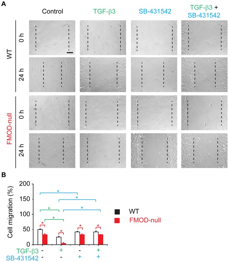
Figure 5. In vitro migration assay of primary dermal fibroblasts derived from adult WT and FMOD-null mice skin.
Cell migration was documented by photographs taken immediately after scraping, as well as 24(A). Migration was quantified by measuring the average wound gap between the wound edges before and after the treatment, and calculated as: Cell migration (%) = (Gap0h-Gap24h)/Gap0h ×100% (B). 100 pM TGF-β3 was used to inhibit dermal fibroblast migration in vitro, while 10 µM TβRI-specific inhibitor SB-431542 was used to block TβRI-mediated signal transduction. Bar = 200 µm. N = 6; *, P<0.05. Red stars indicate the significance that resulted from FMOD-deficiency; green stars indicate the significance that resulted from TGF-β3 application; and blue stars indicate the significance that resulted from SA-431542 blockage of TβRI.

