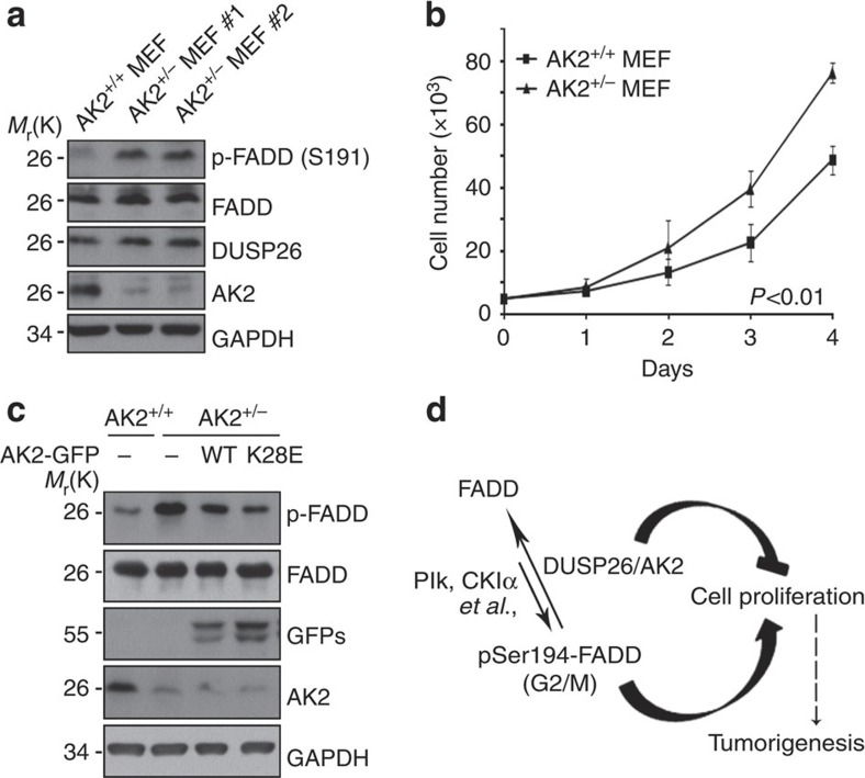
Figure 7. FADD phosphorylation at ser191 and cell proliferation is augmented in AK2+/− MEFs.
(a) Increase of p-FADD (Ser191) in AK2+/− MEFs. On embryonic day 9, MEFs were cultured from wild-type and AK2+/− mouse embryos. Cell extracts were prepared and subjected to western blotting with anti-mouse pSer191-FADD and anti-AK2 antibodies. (b) AK2+/− MEFs show enhanced cell proliferation. Wild-type and AK2+/− MEFs (5 × 10³) were prepared and examined every day for cell proliferation for 4 days. Values are the mean±s.d. (n=3). P<0.01; t-test. (c) Reconstitution of AK2+/− MEFs with AK2 wild-type or AK2 K28E mutant reduces FADD phosphorylation. AK2+/− MEF cells were transfected with wild-type AK2-GFP or AK2 K28E-GFP mutant for 24 h and FADD phosphorylation was examined by immunoblotting. (d) A proposed model of AK2/DUSP26 protein complex in cell proliferation. AK2 forms a protein complex with DUSP26 to regulate FADD dephosphorylation, and thereby cell proliferation.

