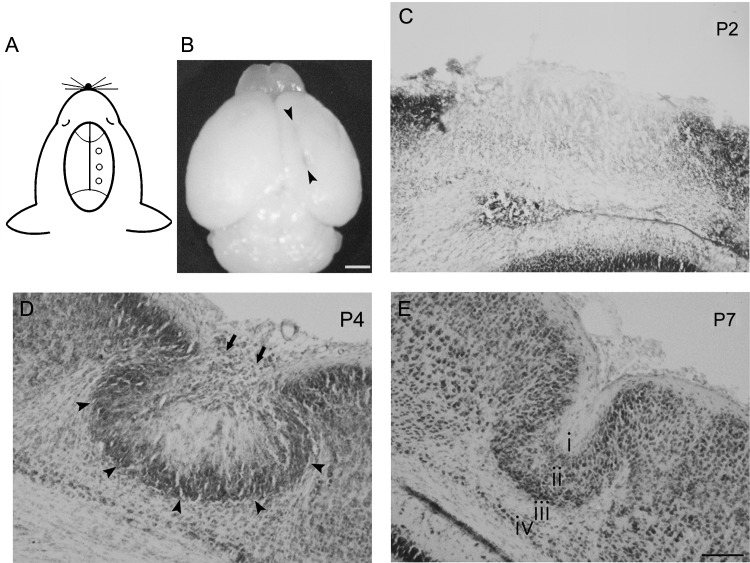
Figure 1.
Morphology of freeze-lesion-induced focal cortical malformations in mice. (A) Schematic illustration indicating the lesioned points in the right hemisphere of a P0 GAD67-GFP KI mouse. (B) Dorsal view of a P7 mouse brain that received the FFL on the day of birth. A longitudinal microgyrus can be observed as an infolding of the brain surface at P7 in the right hemisphere (arrowheads). (C–E) Thionin-stained coronal sections at different stages. At P2, the position of the lesion was identified by necrotic tissue in the lesioned area (C). At P4, a cell-dense layer was observed in the superficial part (arrow) and at the boundaries (arrowheads) of the lesioned area (D). At P7, a microgyric architecture could be detected with a 4-layered (i, layer 1; ii, layer 2; iii, layer 3; iv, layer 4) cortex (E). Scale bars: B, 1 mm; C–E, 100 μm.

