Figure 6.
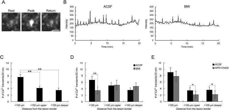
Spontaneous intracellular Ca2+ transients near the lesion site were mediated by tonic GABAA receptor activation. (A) Typical example of a P4 neocortical cell in the area within 100 µm of the lesion border showing spontaneous Ca2+ transients in its soma. (B) Traces of spontaneous Ca2+ transients before and after bath application of 20 µM BMI. (C) Numbers of spontaneous Ca2+ transients in cells in each area. The frequency of Ca2+ transients in the cells within 100 µm of the lesion border (n = 34) was significantly higher than that in the cells in both the upper (n = 34) and the lower (n = 40) parts >100 µm from the lesion border (post hoc Tukey test; **P < 0.01, 15 slices). (D) The number of spontaneous Ca2+ transients before and after bath application of BMI in each area. The frequency of Ca2+ transients was significantly decreased by BMI only in the area within 100 µm of the lesion border (2-tailed paired t-test, **P < 0.01). Number of cells: <100 µm, 21; >100 µm; upper, 21; deeper, 18 (6 slices). (E) Number of spontaneous Ca2+ transients before and after bath application of d-AP5 and CNQX in each area. The frequency of Ca2+ transients was significantly decreased by d-AP5 + CNQX only in the areas >100 µm from the lesion border (paired 2-tailed t-test, *P < 0.05). Number of cells: <100 µm, 13; >100 µm; upper, 13; deeper, 22 (9 slices). Scale bar: 20 μm. ACSF, artificial cerebrospinal fluid control.

