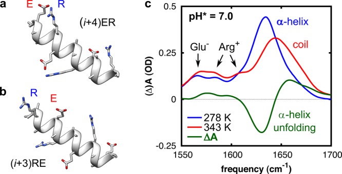
Figure 1.

(a,b) Schematic representation of the folded structure of two of the investigated peptides, showing the salt-bridging side chains Glu– (E) and Arg+ (R). (a) Peptide (i + 4)ER, in which E and R are spaced four peptide units apart. (b) Peptide (i + 3)RE, in which R and E are spaced three peptide units apart and in reverse order. Structures optimized and rendered with Chimera.17 (c) Temperature-dependent Fourier transform infrared (FTIR) spectra of peptide (i + 4)ER. The thermal difference spectrum (green) reflects the conformational changes upon thermal unfolding. The peptide concentration was 12 mM.
