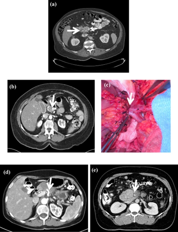
Figure 1.

Representative imaging characteristics of patients with resectable, borderline resectable and locally advanced un-resectable pancreatic cancer. (a) Patient with resectable pancreatic cancer. The bold arrow indicates a hypodense mass. Small arrows indicate the superior mesenteric vein (SMV)/ superior mesenteric artery (SMA), both of which have a clear fat plane around them. (b) A patient with borderline resectable pancreatic cancer. The bold arrow indicates the site where the tumour involves the left side of the portal vein (PV). (c) Intra-operative picture of a borderline resectable tumour involving the PV. This tumour required the resection of the PV. (d) A locally advanced un-resectable tumour encasing the celiac axis depicted by a bold arrow. (e) Locally advanced un-resectable pancreatic cancer encasing the SMA depicted by a bold arrow
