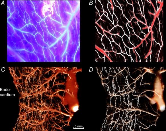
Figure 3.

A, photograph of the epicardial fluorescence (Batson cast material with ultraviolet blue labelling) under excitation with ultraviolet light. B, reconstruction of the same epicardial area after analysis of imaging cryomicrotome data. The automatically detected collateral pathways are depicted in white and the normal vessels in red. C, transmural vessels in a 7.5-mm-thick tissue slab in the longitudinal direction of the heart. D, collateral pathways detected in the same slab are highlighted in white.
