
Figure 4.
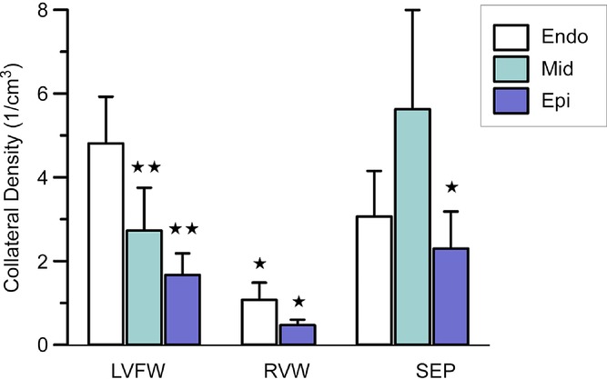
Data are shown for the total number of collaterals obtained from all study hearts. Collateral density was highest in the subendocardium of the left ventricular free wall and mid-myocardium of the septum. Abbreviations: Endo, endocardial layer; Epi, epicardial layer; LVFW, left ventricular free wall; Mid, mid-myocardial layer; RVW, right ventricular wall; and SEP, septum. *P < 0.05 and **P < 0.01, Student's t test compared with the endocardial layer of the LVFW. No significant differences exist between the other groups.