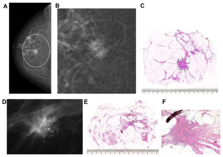
Figure 10.
Right breast, craniocaudal projection (A) and microfocus magnification (B). There is a centrally located solitary stellate malignant tumor with no associated microcalcifications. Large thin-section histology image (C) confirms the 15 × 13 mm unifocal invasive cancer with no extratumoral in situ components. A solitary <10 mm stellate tumor with associated malignant-type microcalcifications (D). Large section (E) and low power histology (F) images demonstrate the single focus invasive cancer associated with low-grade cancer in situ within and outside the invasive tumor.

