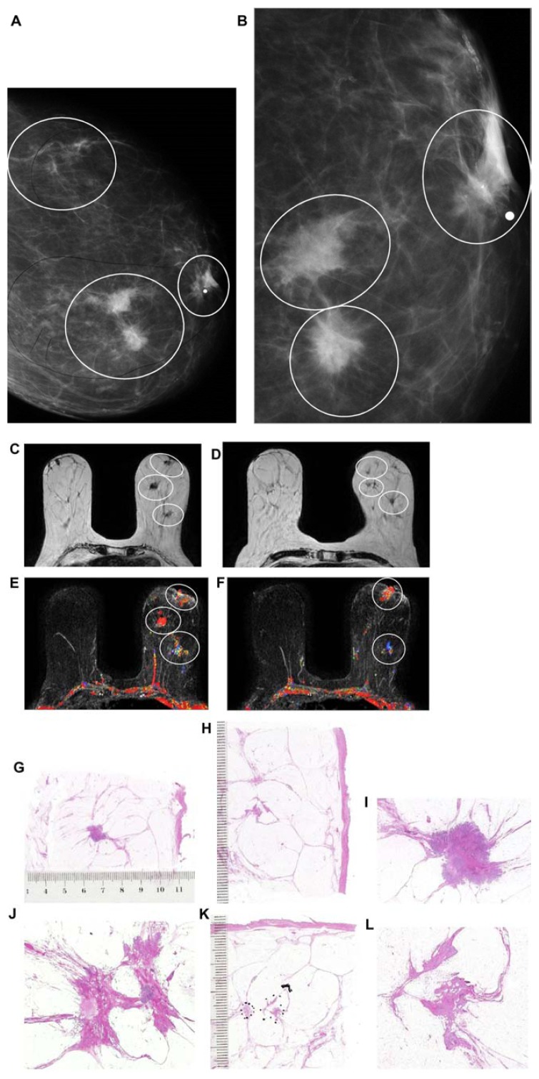
Figure 11.
Left breast mediolateral oblique projection (A) and microfocus magnification (B) shows multifocal stellate malignant tumors with no associated malignant-type calcifications. Breast MRI (C–F) demonstrates the presence of multifocal invasive carcinomas spread over a >10 cm area between the nipple and the chest wall. Multiple large thin-section histology images confirm multifocal (seven foci) invasive lobular carcinoma (the largest focus measuring 11 × 10 mm), ER/PR positive, HER2-negative invasive cancer with low proliferation index (Ki-67: 5%), and no lymph vessel invasion (LVI). Histologic examination of 16 surgically removed axillary lymph nodes showed no metastases (pN0/16).

