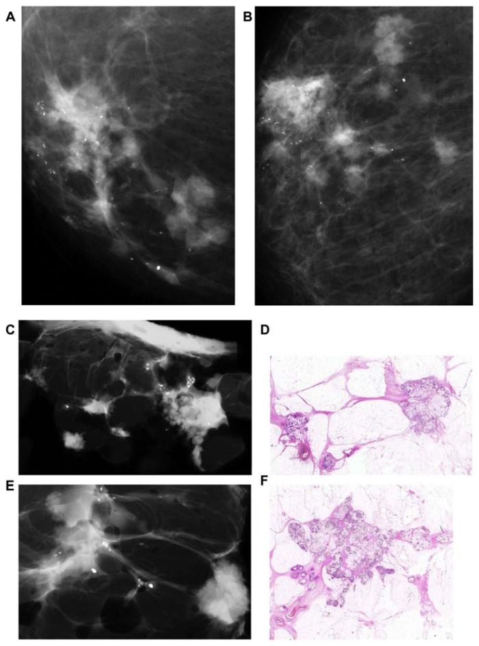
Figure 12.
Microfocus magnification images on the craniocaudal and the lateromedial horizontal projections (A,B) demonstrating ill-defined circular lesions bridged together, associated with crushed stone-like, malignant-type calcifications. Mastectomy specimen slice radiographs (C,E) and large thin-section histology images confirm the presence of 20 invasive mucinous cancer foci (the largest focus measuring 15 × 15 mm) associated with 65 × 40 mm Grade 2 in situ carcinoma, pNX. LVI was present.

