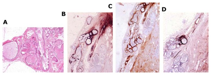
Figure 18.
Large thin-section histology image (A) shows abnormal, contorted ducts with “micropapillary cancer in situ.” The accumulated fluid distends the ducts and branches. Tenascin staining (B–D) demonstrates overexpression, indicating that the torturous, duct-like structures correspond to neoductgenesis.

