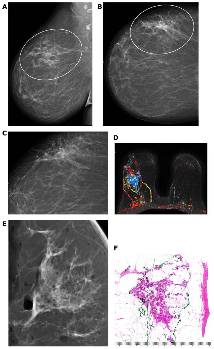
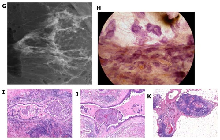
Figure 23.
A–K Mammographically detected architectural distortion in the upper-outer quadrant of the right breast with no associated microcalcifications. There is also a pathologic lymph node in the right axilla (A,B). Microfocus magnification (C) and breast MRI (D) demonstrate the large extent of the disease. Specimen slice radiographs (E,G) compared with large thin-section (F) and large thick-section histology (H) images show the cancer-filled, tortuous, newly formed ducts tightly packed together. Low power histology images (I,J,K) demonstrate that the abnormal, cancer-filled, duct-like structures with micropapillary cell architecture are surrounded by a thick desmoplastic reaction and lymphocytic infiltration, which are the histologic signs of neoductgenesis.


