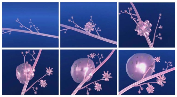
Figure 6.
(A–F) Computer simulation images of the development of Grade 2 in situ carcinoma within the TDLU. The lobule gradually becomes distended and deformed. Calcifications are formed within the necrotic debris and are seen on the mammogram as crushed stone-like/pleomorphic calcifications. (Artwork from Ref. 8. Copyright by Georg Thieme Verlag. Used with permission).

