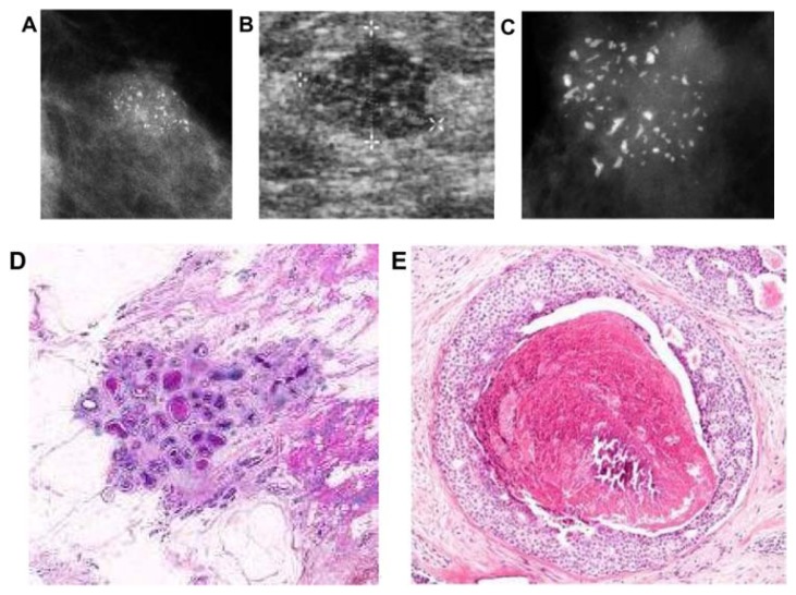
Figure 7.
(A) A solitary cluster of crushed stone-like (pleomorphic) calcifications was detected at mammography screening. (B) Ultrasound examination: a 12-mm solitary hypoechogenic area with calcifications, corresponding to a distended TDLU. (C) Microfocus magnification specimen radiograph: malignant-type calcifications in a cluster. (D) Low power large section histology image of the solitary TDLU, distended and distorted by the Grade 2 in situ carcinoma. (E) Higher power magnification of a single acinus: solid and cribriform cancer in situ with central necrosis and amorphous calcification distending the acinus. Histologic diagnosis: Grade 2 in situ carcinoma within the TDLU (unifocal AAB).

