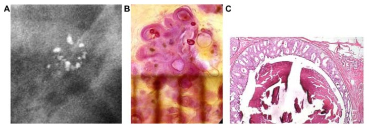
Figure 8.
(A) Mammographically detected solitary cluster of crushed stone-like calcifications with no associated tumor mass. (B) Thick-section (3D) histology image: the individual acinus is distended and each measures approximately 1 mm (which corresponds to the size of an entire normal TDLU).This remarkable growth is the result of increased intraluminal pressure because of accumulation of cancer cells, intraluminal necrosis, and amorphous calcification. (C) Low power histology image of a segment of one distended acinus showing cribriform cell-architecture, intraluminal necrosis, and a central amorphous calcification. Histologic diagnosis: Grade 2 in situ carcinoma localized to a single TDLU. Note that the malignant process is confined to a TDLU and is not within the ducts, so that it is not “ductal” carcinoma in situ (DCIS).

