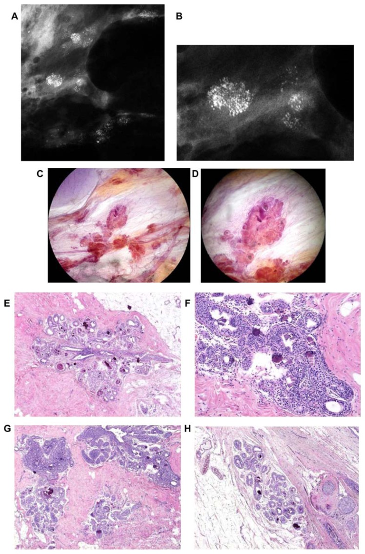
Figure 9.
Screen-detected multiple clusters of powdery, dust-like calcifications; specimen radiograph with and without microfocus magnification (A,B). Thick-section (3D) histology images (C,D) demonstrate that the calcifications are localized within distended TDLUs. Low (E,G,H) and intermediate (F) power histology images: low-grade cancer in situ localized to the TDLUs. The powdery calcifications seen on the mammograms correspond to psammoma body-like calcifications. Histology: Grade 1 in situ carcinoma localized to the TDLU (not “ductal” carcinoma in situ).

