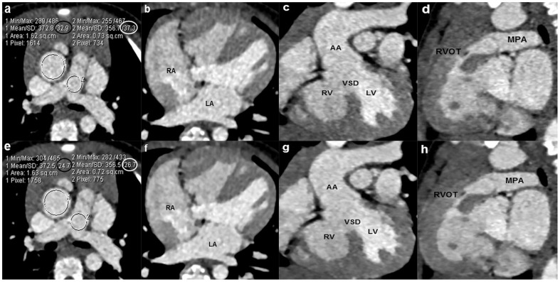
Figure 1. An 18-month boy with the diagnosis of Tetralogy of Fallot and atrial septal defect.
The sequential DSCT angiography was performed with 70/rotation (effective radiation dose, 0.32 mSv). Axial images (a,e), multiplanar reformatted (MPR) images (b–d, f–h) using filtered back projection (FBP) algorithm (a–d) and sinogram affirmed iterative reconstruction (SAFIRE) algorithm (e–h) are shown. Image noise of the ascending aorta and pulmonary trunk which is expressed as the standard deviation (SD) of the attenuation (HU) in the regions of interest is significantly reduced in images reconstructed by SAFIRE (black and white circles in f) in contrast to FBP black and white circles in a). MPR images reconstructed with SAFIRE algorithm exhibit substantially reduced image noise and the improved image quality compared with images obtained with FBP. LA = left atrium, RA = right atrium, RV = right ventricle, LV = left ventricle, VSD = ventricular septal defect, AA = ascending aorta, RVOT = right ventricular outflow tract, MPA = main pulmonary artery.

