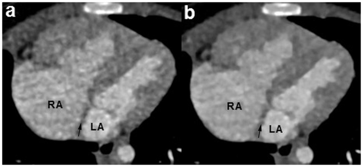
Figure 2. A 7-month girl with a small atrial septal defect.

The sequential DSCT angiography was performed with 70/rotation (effective radiation dose, 0.21 mSv). Multiplanar reformatted (MPR) image reconstructed with sinogram affirmed iterative reconstruction (SAFIRE) algorithm (b) shows the small atrial septal defect (arrow) clearly. However, the lesion was missed on the MPR image using filtered back projection (FBP) algorithm (a). LA = left atrium, RA = right atrium.
