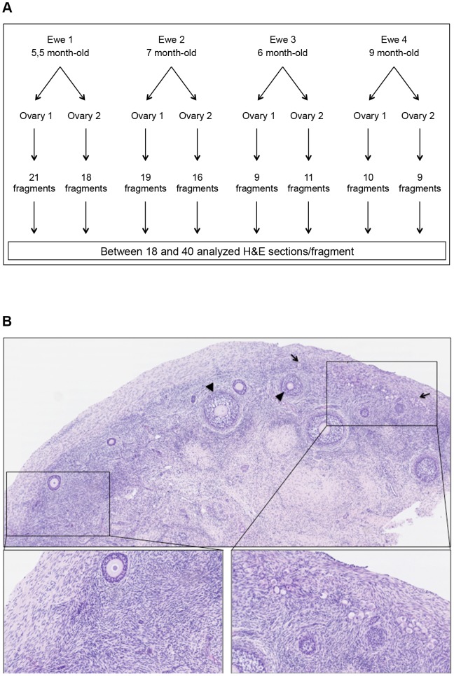
Figure 1. Flow chart of the experimental design (A) and representative histology of a sheep cortex section (B).
Ovaries were harvested from two ewes and fully cut into cortical fragments. Subsequently, each fragment was serially and completely sectioned, and approximately 40 H&E sections, each 30 µm apart from one another, were further used for the follicular quantification (A). The uneven repartition of follicles within the sheep ovarian cortex is obvious (B). The left part of the H&E section is completely devoid of primordial follicles, whereas the right part contains mostly primordial follicles. Primordial follicles (plain arrows) and secondary follicles (arrowhead).

