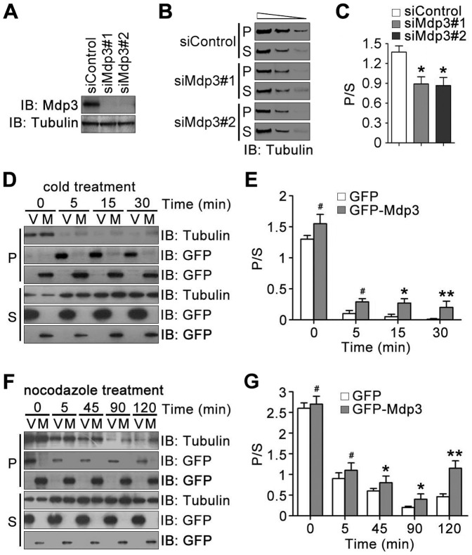
Figure 1. Mdp3 protects microtubules from cold- or nocodazole-induced depolymerization.
(A) HeLa cells were transfected with control or Mdp3 siRNAs. The levels of Mdp3 and α-tubulin were then examined by immunoblotting with antibodies against Mdp3 and α-tubulin. (B) HeLa cells were transfected with control or Mdp3 siRNAs, placed on ice for 30 min, and then re-incubated at 37°C for 30 min to permit microtubule regrowth. Tubulin partitioning between the polymer (P) and soluble dimer (S) forms was examined by immunoblotting with the anti-α-tubulin antibody. Each of the polymeric and soluble fractions was loaded at a 3-fold serial dilution to ensure the intensity of bands on the blots in the linear range of detection. (C) Experiments were performed as in (B), and the ratio of tubulin polymer to soluble dimer was analyzed. (D) HeLa cells were transfected with GFP (V) or GFP-Mdp3 (M) and placed on ice for 0, 5, 15, or 30 min. Tubulin partitioning between the polymer and soluble dimer forms was examined by immunoblotting with the anti-α-tubulin antibody, and the expression of GFP and GFP-Mdp3 was examined by immunoblotting with the anti-GFP antibody. (E) Experiments were performed as in (D), and the ratio of tubulin polymer to soluble dimer was analyzed. (F) HeLa cells were transfected with GFP or GFP-Mdp3 and treated with nocodazole for 0, 5, 45, 90, or 120 min. Tubulin partitioning between the polymer and soluble dimer forms was examined by immunoblotting with the anti-α-tubulin antibody, and the expression of GFP and GFP-Mdp3 was examined by immunoblotting with the anti-GFP antibody. (G) Experiments were performed as in (F), and the ratio of tubulin polymer to soluble dimer was analyzed. *, p<0.05; **, p<0.01; #, not significant (p≥0.05).

