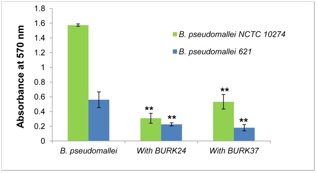
Figure 7. Quantification of biofilm formation in absence and presence BURK24 and BURK37.
5×10⁵ CFU/ml of B. pseudomallei NCTC 10274 and B. pseudomallei strain 621 were separately suspended in TSB with 50 mM glucose containing respective bacteriostatic concentration of BURK24 and BURK37 and incubated for 24 h at 37°C. Formation of biofilm was quantified with crystal violet stain. Both mAbs showed varied but considerable percentage of inhibition on biofilm formation capability of the strains studied. The graphical data presented here is average (n = 6) of A570 nm of the experiment. The error bar represents standard error.

