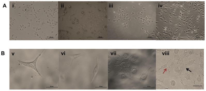
Figure 10. Morphological study on effect of mAbs on pathogen induced apoptosis using A549 cells.
Phase contrast image of A549 cells in (A) 10× and (B) 40× magnification. (i and v) Phase contrast image of uninfected A549 control cells showing intact cell morphology. (ii and vi) Phase contrast image of A549 cells treated with B. pseudomallei NCTC 10274 for a dutration of 24 h in presence of BURK24. (iii and vii) A549 cells treated with B. pseudomallei NCTC 10274 in presence of BURK37. (iv and viii) A549 cells infected with B. pseudomallei NCTC 10274 showing apoptotic morphologies including reduction in cell volume and rupturing of cell membrane upon 24 h post-infection. (viii) Red arrow shows cell fusion and black arrow shows rupturing of cell membrane.

