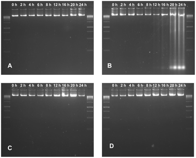
Figure 11. DNA damage assay of A549 cells.
DNA damage assay was performed to study the pathogen induced apoptosis, where A549 cells were infected with B. pseudomallei NCTC 10274 in presence and absence of bacteriostatic concentration of BURK24 and BURK37. In a duration of 24 h, A549 cells were permeabilized and DNA was extracted at intervals of 0 h, 2 h, 4 h, 6 h, 8 h, 12 h, 16 h, 20 h and 24 h (A) DNA of A549 cells uninfected with B. pseudomallei (B) DNA of A549 cells infected with B. pseudomallei. DNA damage is noticed after 16 h of infection. (C) and (D) DNA of A549 cells infected with B. pseudomallei in presence of 30 µg/ml of BURK24 and 62.5 µg/ml of BURK37 respectively. The intactness of DNA in case of A549 cells treated with the pathogen in presence of mAbs is comparable to that of cells untreated with bacteria, inferring the efficiency of BURK24 and BURK37 to prevent apoptosis.

