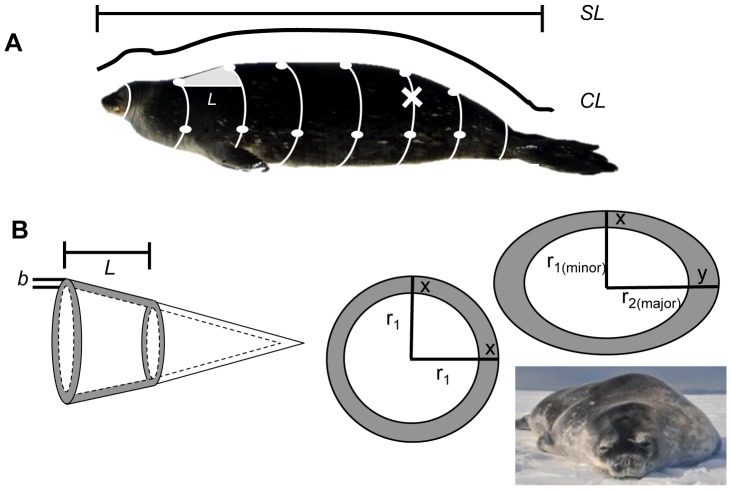
Figure 2. Morphometric measurements taken for each study animal.
(A) SL = Standard length, CL = Curve length, Girths = white lines, Blubber depths = white dots, Cone section length calculations = grey triangle and “L”. Site of blubber biopsy is marked with “X”. (B) Reconstruction of truncated cones with segment length “L” and blubber depth “b” (At left). Circular and elliptical cross-sections shown (At right). Because an ellipse has a major and minor radius “r,” the model can account for different dorsal and lateral blubber depths (x and y) and more accurately reflect true animal shape.

