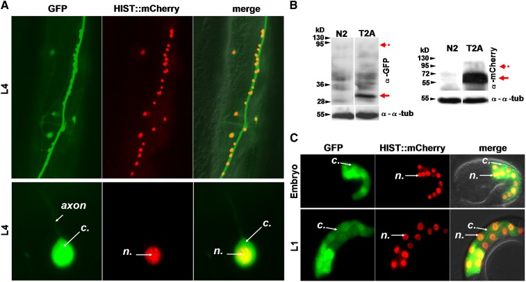
Figure 3.
2A viral peptides allow the efficient production of two independent polypeptides in different tissues. (A) Localization pattern of GFP and HISTONE::mCherry in neuronal cells in transgenic lines expressing a F25B3.3p::GFP::T2A::histone::mCherry construct. GFP localizes within the whole cell whereas HISTONE-mCherry remains tightly restricted to the nucleus. n, nuclear; c, cytosol. The ventral midbody area of the worm is shown. The worm is oriented vertically with its tail to the bottom. (B) Western blot analysis showing the production of independent GFP and mCherry proteins from a T2A construct in neuronal cells. The GFP and HISTONE-mCherry products, detected at 30 and 60 kDa, respectively, are indicated by red arrows; dotted arrows indicate the expected size for an uncleaved product; α-tubulin was used as a loading control. (C) Localization pattern of GFP and HISTONE::mCherry in intestinal cells of embryos and L1 larvae in lines expressing a ugt-22p::GFP::T2A::histone::mCherry construct. GFP localizes within the whole cell whereas HISTONE-mCherry remains tightly restricted to the nucleus. n, nuclear; c, cytosol. The midbody area of L1 larvae oriented vertically with the tail to the bottom is observed. See Table S3 for information on the different transgenic lines obtained.

