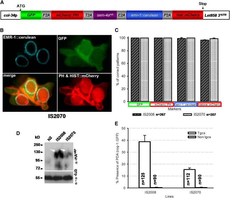
Figure 4.
2A viral peptide technology allows simultaneous delivery of multiple independent and functional polypeptides in the worm. (A) Schematic view of the molecular construct encompassing five products fused in frame with F2A, T2A, E2A, and P2A. GFP is addressed to the whole cell, mCherry::PH (Pleckstrin Homology domain) is addressed to the cellular membrane, EMR-1::cerulean (Ce-emerin) is addressed to nuclear membrane (Lee et al. 2000), and HISTONE::mCherry is addressed to the nucleus. col-34p was used to drive expression in the rectal cells (Kagias et al. 2012). (B) Confocal imaging of rectal cells of early L1s expressing col-34p::GFP::F2A::mCherry-PH::T2A::sem-4A-HA::E2A::emr-1::cerulean::P2A::histone::mCherry. All the markers are addressed to the expected cellular compartments (see also Figure S5); the Y, B, and F rectal cells are shown; anterior is to the left and ventral to the bottom. (C) Quantification of the presence of all five markers at the correct compartment. We noted in one of the two lines (IS2008) that the fifth protein, HISTONE::mCherry, is not seen in 3.3% of the cells scored. n, total number of cells scored. (D) Western blot analysis showing the presence of SEM-4a::HA as one band at ∼100 kDa as expected (white arrow) in the two transgenic independent lines IS2008 and IS2070. (E) Rescuing efficiency of the sem-4(n1971) “no PDA” defect in the two independent lines IS2008 and IS2070 expressing col-34p::GFP::F2A::mCherry-PH::T2A::sem-4A-HA::E2A::emr-1::cerulean::P2A::histone::mCherry. Interestingly, the rescuing efficiency for each line correlated with the levels of expression of SEM-4a-HA detected by Western Blot. For each experiment, nontransgenic (Non tgcs) siblings were 100% PDA defective. n, number of animals scored.

