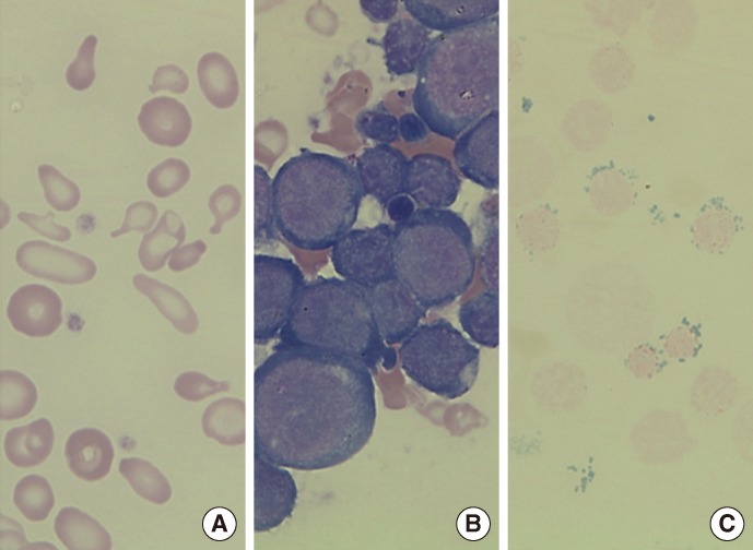
Fig. 1.
(A) Peripheral blood smear showing dimorphic red blood cells (RBCs) including microcytic hypochromic RBCs with severe anisopoikilocytosis (Wright-Giemsa stain, ×1,000). (B) Bone marrow (BM) aspirate smear showing marked increase of erythroid precursors (Wright-Giemsa stain, ×1,000). (C) Iron stain of BM aspirate showing many ringed sideroblasts, which accounted for up to 50% of the total erythroid precursors (Prussian blue stain, ×1,000).

