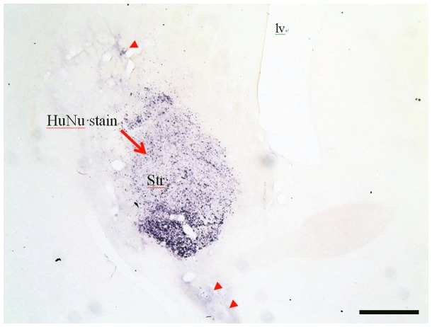
Figure 4. A representative image of HuNu staining in the striatum.
HuNu+cells were confined to the striatum of rats receiving the lowest dose of graft cells (Group B). Note that small amounts of HuNu staining extended both dorsally and ventrally from the striatum. Scale bar = 0.5 mm [Iv; lateral ventricle, Str: striatum].

