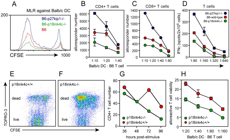
Figure 4. Effect of CDK inhibitor deficiency on in vitro alloimmune responses.
CFSE-labeled lymphocytes from B6 WT (red symbols), p27kip1−/− (blue symbols), or p18ink4c−/− (green symbols) mice were cultured with irradiated DC derived from BALB/c bone marrow at the indicated T cell-to-DC ratios for 5 days. Alloresponsive cells were identified by dilution of CFSE (A), and the alloresponsive CD4+ T cells (B) and CD8+ T cells (C) were enumerated by flow cytometry using reference beads. The frequency of allospecific IFNγ-producing cells (D) was assessed at day 3 by replating responders and BALB/c DC in ELISPOT cultures overnight. CD4+ T cell viability was assessed by flow cytometry using the vital dye TOPRO-3 (34) in cultures stimulated in vitro with anti-CD3 Ab (E–G) or with allogeneic DC (H). Data are plotted as the mean +/−SEM of duplicate cultures, and are representative of 2–3 independent experiments.

