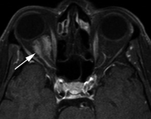
Fig. 1.

Post-contrast axial T1-weighted sequence in a 64-year-old man with proptosis demonstrates a homogeneously enhancing, intraconal mass within the right orbit (arrow) that partially encircles the right optic nerve. Smooth dural thickening and enhancement extends posteriorly to the optic canal. The imaging appearance is consistent with a meningioma
