
Fig. 10.
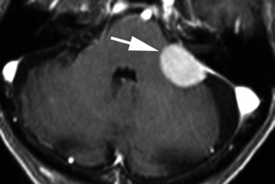
Post-contrast axial T1-weighted sequence in an 83-year-old man with dizziness and falls demonstrates a homogeneously enhancing, extra-axial mass within the left cerebellopontine angle (arrow) with a broad dural attachment and a dural tail extending anterior to the left internal acoustic meatus and posteriorly to the sigmoid sinus. The mass overlies the left internal acoustic meatus without intracanalicular extension. Appearance and histology are consistent with a meningioma (grade I)