
Fig. 12.
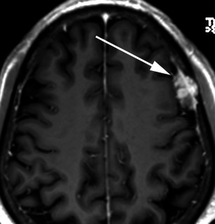
Post-contrast axial T1-weighted MRI in a 73-year-old man with headaches demonstrates a mildly heterogeneously enhancing extra-axial mass over the left lateral convexity (arrow) consistent with a meningioma at histology that invades the inner table and diploic space of the left frontal bone. The outer table is contacted and thinned without breach