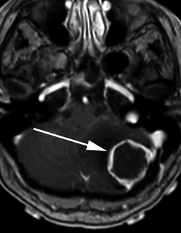
Fig. 17.

Post-contrast axial T1-weighted sequence in a 68-year-old woman with headache and ataxia demonstrates a ring enhancing mass within the left posterior cranial fossa (arrow) with a broad dural base posterolaterally. There is surrounding vasogenic oedema with midline shift and effacement of the fourth ventricle. Histology at surgery was a meningioma with central necrosis secondary to infarction
